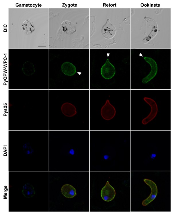
Figure 3.
Localization of PyCPW-WPC-1 in sexual-stage parasites. Using an immunofluorescent assay (IFA), PyCPW-WPC-1 was found to be localized to the surface of developing ookinetes, including zygotes, retorts, and mature ookinetes (shown in green). The surface of developing ookinetes was stained with antibodies targeting Pys25 (red), a well-known ookinete surface protein. Nuclei were stained by DAPI (blue). The merged images represented the colocalization of PyCPW-WPC-1 and Pys25 on the parasite surface. Parasite morphology was analysed by differential interference contrast (DIC) imaging. The bar indicates 5 μm.

