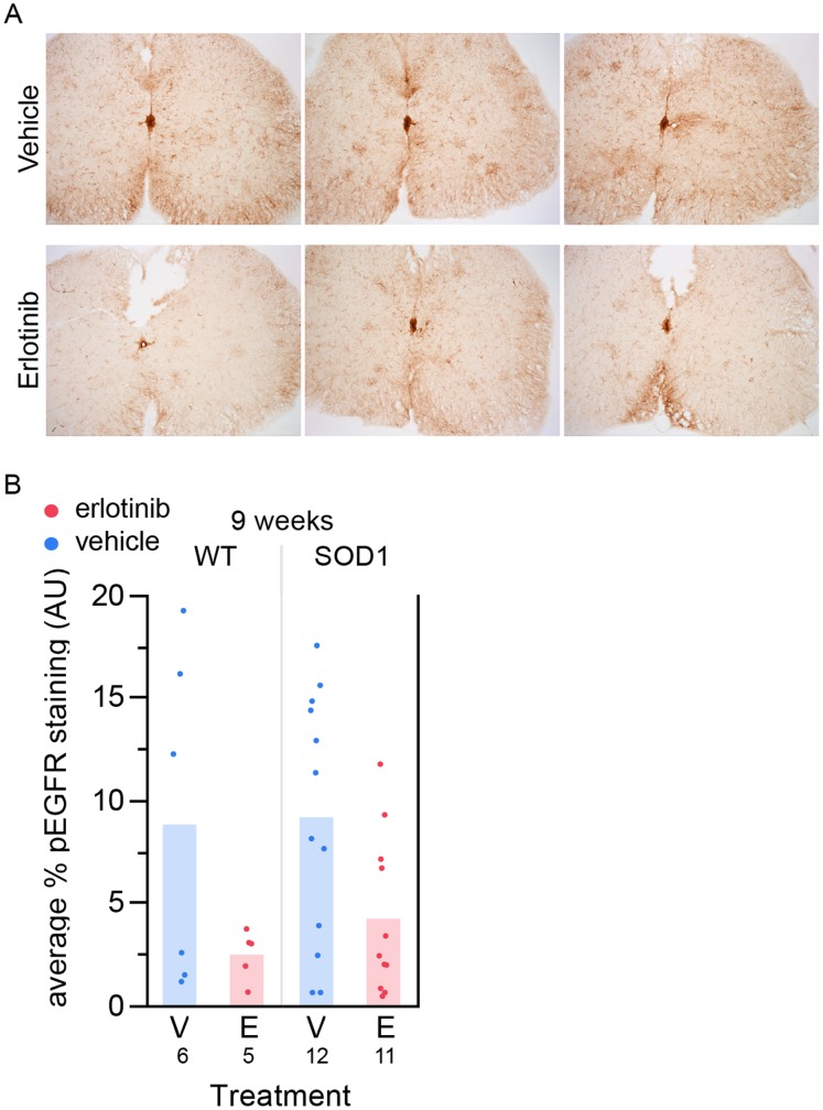
Figure 5. Erlotinib reduces anti-pEGFR staining in spinal cord.
(A) Sample images from 6 different mice of anti-pEGFR staining in spinal cord. Top 3 images: vehicle-treated; bottom 3 images: erlotinib-treated. These images are not extreme examples from either group; they are representative of average levels of staining for each treatment group. (B) Quantification of staining using the Novus anti-pEGFR antibody in the 9-week lumbar spinal cord tissue, 24 hours post-last dose. Erlotinib significantly reduces the staining. p = 0.0124, one-tailed t test. The n/group is listed below each bar. V – vehicle; E – erlotinib.

