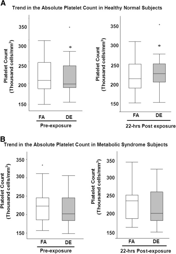
Figure 1.
Absolute Platelet Count in Healthy Normal Subjects (A) and Metabolic Syndrome Subjects (B) following exposure. Figure showing the distribution of the platelet count at the different time points in (A) Healthy subjects and (B) Metabolic Syndrome subjects. Each panel depicts medians (bars) and interquartile ranges (whiskers) for platelet count associated with each exposure at baseline (pre-exposure) and 22 hours post exposure initiation. Crosses above the panels represent those that are outside of the interquartile range; * indicates P=0.04 for the difference between DE pre-exposure and 22hours post exposure in healthy normal subjects. FA=filtered air; DE=200 μg/m3 (PM2.5) DE.

