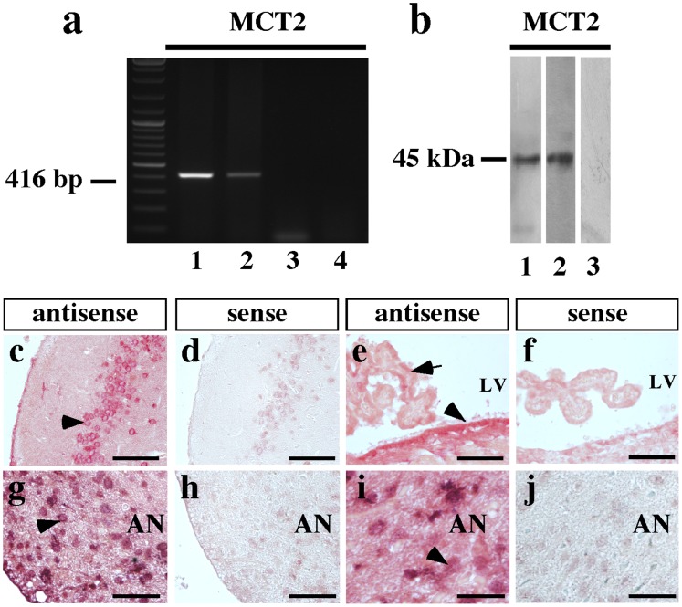
Figure 6. MCT2 expression in adult rat hypothalamus.
(a) RT-PCR analysis of RNA isolated from rat cerebral cortex (lane 1) and rat hypothalamus (lanes 2–3). RT(−) (lane 3) and water in the PCR reaction (lane 4). (b) Immunoblot analysis of total protein extracted from rat cerebral cortex (lane 1) and rat hypothalamus (lane 2). Negative control was performed with primary antibodies pre-adsorbed with inductor peptide (lane 3). (c–h) Neuronal MCT2 mRNA detection by in situ hybridization. Frontal section of rat brain probed with a MCT2 antisense riboprobe. A representative panoramic image showing high hybridization signal in basal hypothalamus (c), in neurons of entorrinal cortex (d). Ependymal cells of lateral ventricle and some choroidal cells present positive hybridization with antisense MCT2 riboprobe (e). In the hypothalamus, a high hybridization signal was observed in neurons of peri-ventricular (g) and distal (i) arcuate nucleus. Negative reaction was obtained using sense riboprobes in control areas (insets in c–g). AN: arcuate nucleus, LV: lateral ventricle. Scale bar: (c–f) 50 µm; (g–j) 20 µm.

