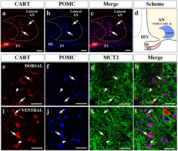
Figure 11. MCT2 is localized in anorexigenic neurons of AN.
(a–c) Low magnification images of ventro-medial hypothalamus, analyzed by immunohistochemistry using anti-CART (red) and anti-POMC (blue) antibodies. Lateral arcuate nucleus neurons show a segregated distribution of both neuropeptides (c). (d) Scheme summarizing the localization of the anorexigenic neuropeptides in the AN. (e–l) High magnification images showing a dorsal (e–h) and ventral (i–l) region of ventricular AN. MCT2 immunoreaction was detected in some neuronal bodies that express one or both anorexigenic neuropeptides, in both areas (h and l). AN: arcuate nucleus, IIIV: third ventricle, ME: median eminence, PT: pars tuberalis. Scale bars: (a–c) 150 µm; (e–l) 50 µm.

