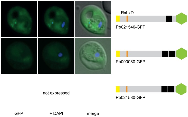
Figure 2. RxLxD containing proteins Pb021540-GFP and Pb000080-GFP are exported into the RBC cytosol.
Pb021580-GFP was not expressed in two independent transgenic parasite lines. GFP fluorescence is indicated by GFP (green), parasite nuclei are stained with DAPI (blue) and merged images include the bright field. A schematic structure of all GFP chimera is depicted beside the panel: signal peptide/hydrophobic stretch (yellow), PEXEL (orange), predicted TMD region (black) and GFP (green).

