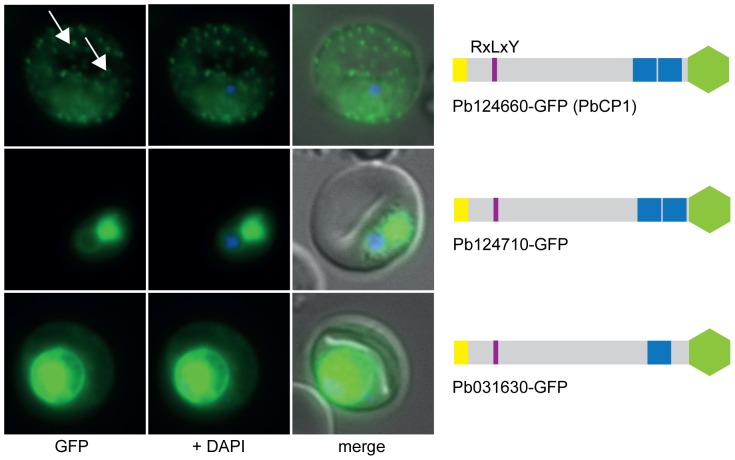
Figure 3. All RxLxY containing proteins are exported into the RBC.
Lower expression levels in the RBC cytosol and accumulation of GFP chimera at the ER and/or PV(M) could also be observed for Pb124710-GFP and Pb031630-GFP. Pb124660-GFP (PbCP1) was also found to localise to extra-parasitic structures within the RBC cytosol (white arrows). GFP fluorescence is indicated by GFP (green), parasite nuclei are stained with DAPI (blue) and merged images include the bright field. A schematic structure of all GFP chimera is depicted beside the panel: signal peptide/hydrophobic stretch (yellow), PEXEL (purple), predicted TMD region (blue) and GFP (green).

