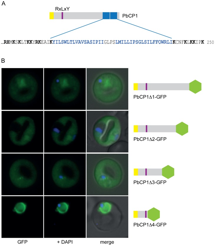
Figure 5. Dissecting the trafficking requirements of PbCP1.
(A) Extract of the PbCP1 C-terminus depicting the predicted 2TMD region (blue and bold), which is flanked by several positively charged residues (black and bold). (B) Consecutive 50aa truncations of PbCP1 were undertaken and the localisation of the respective GFP chimera analysed in live PbANKA parasites. Deletion of the 2TMD region abolished export to the punctuate structures in the respective PbCP1Δ1-GFP chimera, but export into the RBC cytosol was unaffected. Further truncations of 100aa (PbCP1Δ2-GFP) and 150aa (PbCP1Δ3-GFP), respectively, had no effect on export of the corresponding GFP fusion proteins. In the 200aa deletion, GFP is fused directly to the PEXEL motif and led to the accumulation of PbCP1Δ4-GFP within the parasite and the parasite periphery.

