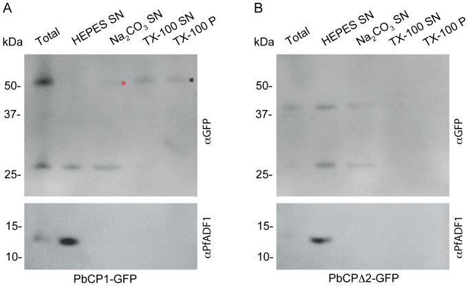
Figure 9. PbCP1-GFP is a membrane-bound protein.
(A) PbCP1-GFP mainly associates with the Triton X-100 soluble (TX-100 SN) and insoluble pellet fractions (TX-100 P) as indicated by an ∼52 kDa protein band (black *) in Western blot analysis. A small fraction is also detected in the supernatant (SN) after Na2CO3 extraction (red *). (B) The PbCP1Δ2-GFP mutant lacking the 2TMD is soluble. Cross-reactive PfADF1 antibodies detected the soluble protein exclusively in the supernatant after hypotonic lysis at the predicted MW of ∼13 kDa. The ∼27 kDa protein band is indicative of cleaved GFP, which no longer associates with the Triton X-100 soluble or insoluble fractions.

