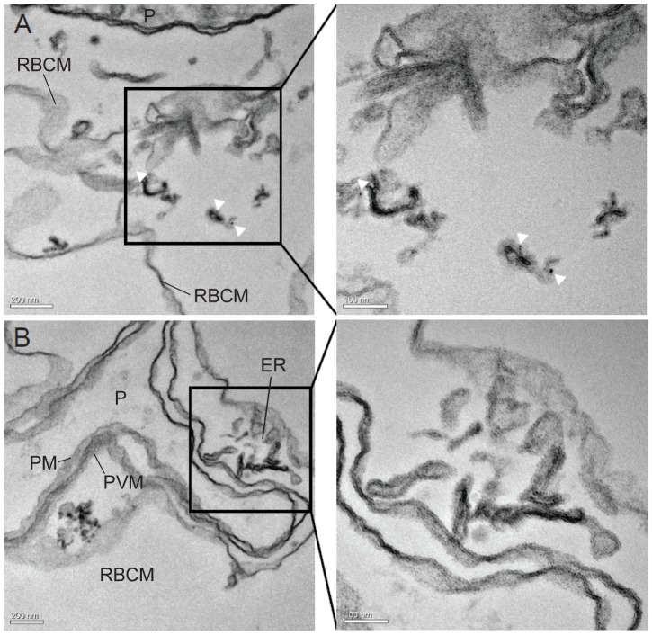
Figure 10. PbCP1-GFP localises to the parasite-induced structures.
(A) IEM was performed on the PbCP1-GFP expressing cell line to localise the protein at the ultrastructural level. Anti-GFP conjugated gold particles decorate the extra-parasitic structures (white arrow heads), indicating that PbCP1-GFP associates with these parasite-induced membranous compartments. (B) In contrast, no labeling of the erythrocytic endoplasmatic reticulum (ER) could be detected. Enlargement of the regions boxed in the panels A and B are shown to their right. The parasite (P), parasite plasma membrane (PM), the parasitophorous vacuole membrane (PVM and the red blood cell membrane (RBCM) are indicated.

