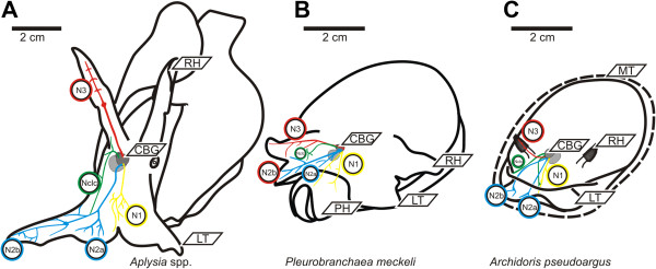
Figure 2.

Neuroanatomy. A: Neuroanatomical scheme of the four cerebral nerves of the investigated Aplysiomorpha, the two investigated Aplysia show no significant differences (only right hemisphere shown). B: Neuroanatomical scheme of the four cerebral nerves of the investigated Pleurobranchomorpha Pleurobranchaea meckeli (only right hemisphere shown). C: Neuroanatomical scheme of the four cerebral nerves of Archidoris pseudoargus (only right hemisphere shown). Abbreviations (referring to all subfigures): CBG – cerebral ganglia, LT – labial tentacle, N1 in yellow, N2 (N2a= inner branch, N2b= outer branch) in blue, N3 in red and the Nclc in green; OV – oral veil, RH – rhinophore.
