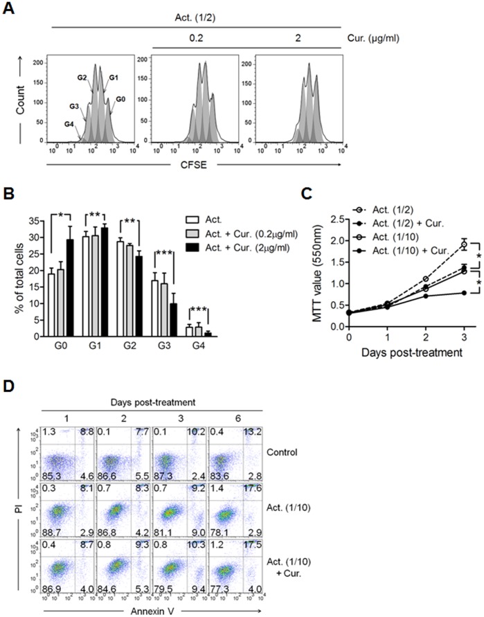
Figure 1. Curcumin inhibits CD4+ T cell expansion induced by CD2/CD3/CD28 signaling without inducing cell death.
CD4+ T cells were cultured in the presence of anti-CD2/CD3/CD28 antibody-coated beads only (Act.) or with either 0.2 or 2 µg/mL of curcumin (Cur.) for the indicated time periods. Act. (1/2) and Act. (1/10) represent bead-to-cell ratios of 1∶2 and 1∶10, respectively. (A, B) For cell proliferation assay, the cells were labeled with CFSE prior to culture, and harvested at 3 days of culture. (A) Cell generations (G0∼G4) were calculated and (B) plotted as the percentage of total cells by using flow cytometry and FlowJo software. (C) Results of an MTT cell proliferation assay to assess cell numbers at 1, 2 and 3 days of culture. (D) Cells were harvested at the indicated time points and labeled with an anti-Annexin V antibody and PI. The numbers in the plot indicate the percentage of cells in the respective areas. Data are representative of 3 experiments yielding similar results. (C and D) Curcumin was added at a concentration of 2 µg/mL. (B and C) Data are presented as the mean ± SD. *P<0.05, **P<0.01, ***P<0.001.

