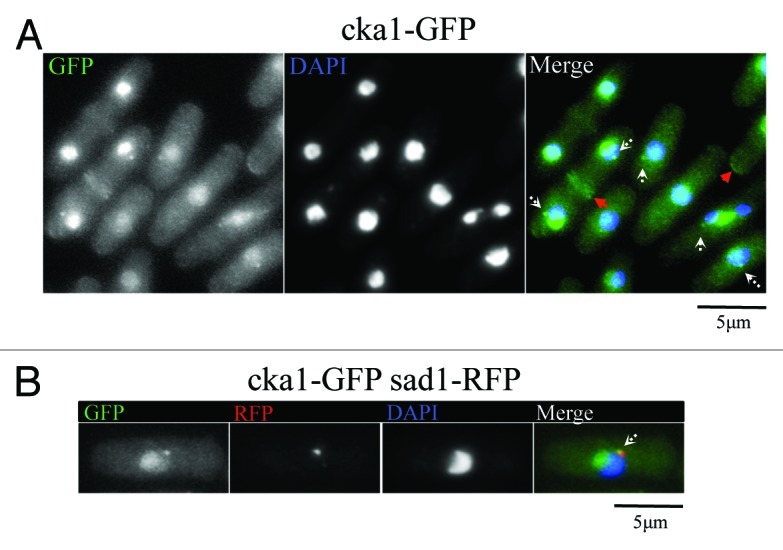
Figure 2. Cka1-GFP localizes similar to MOR pathway proteins like Nak1. (A) Fixed Cka1-GFP-expressing cells (YDM2969) were stained with DAPI. White arrows in the merge panel indicate localization of Cka1-GFP to dots that might correspond to the SPB. Red arrows indicate Cka1-GFP localization to cell septum and tips. (B) Cka1 co-localizes with the SPB marker Sad1. Fixed Cka1-GFP Sad1-RFP expressing cells (YDM3463) were stained with DAPI.
