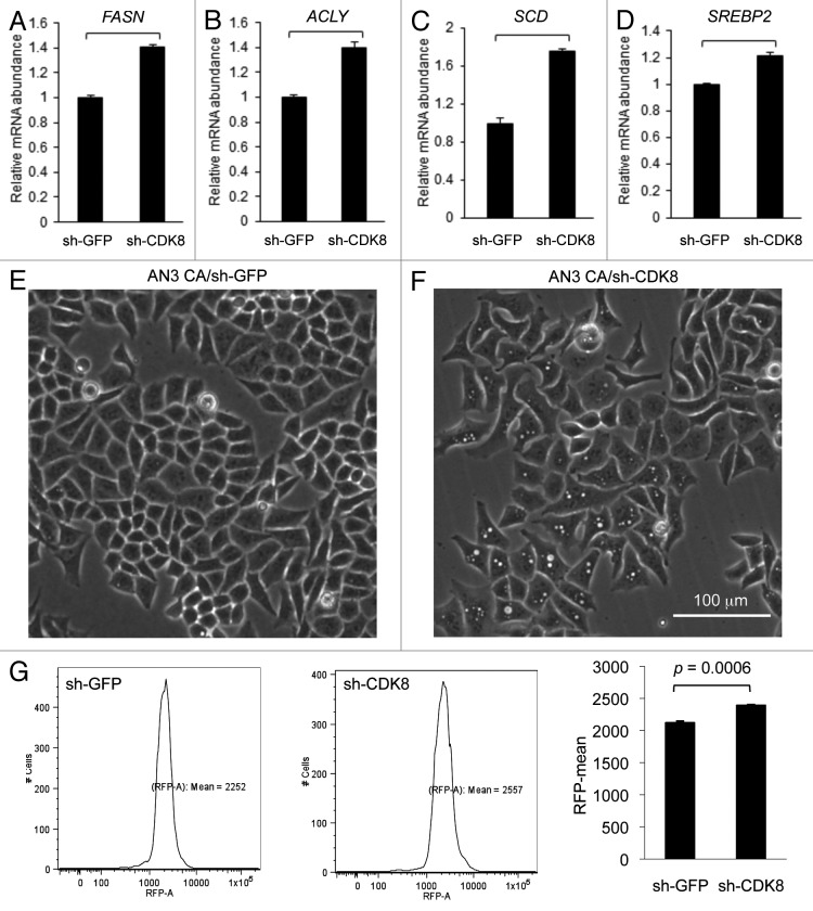
Figure 6. Depleting CDK8 enhances lipid accumulation in endometrial cancer AN3 CA cells. (A–D) Quantification of gene expression by using qRT-PCR. (E–F) The phase contrast images were acquired, showing lipid droplet accumulation in cells with depleted CDK8 (F) compared with the control (E). (G) Cells were stained with Nile Red and subjected to flow cytometry assays to quantify the lipid levels.

An official website of the United States government
Here's how you know
Official websites use .gov
A
.gov website belongs to an official
government organization in the United States.
Secure .gov websites use HTTPS
A lock (
) or https:// means you've safely
connected to the .gov website. Share sensitive
information only on official, secure websites.
