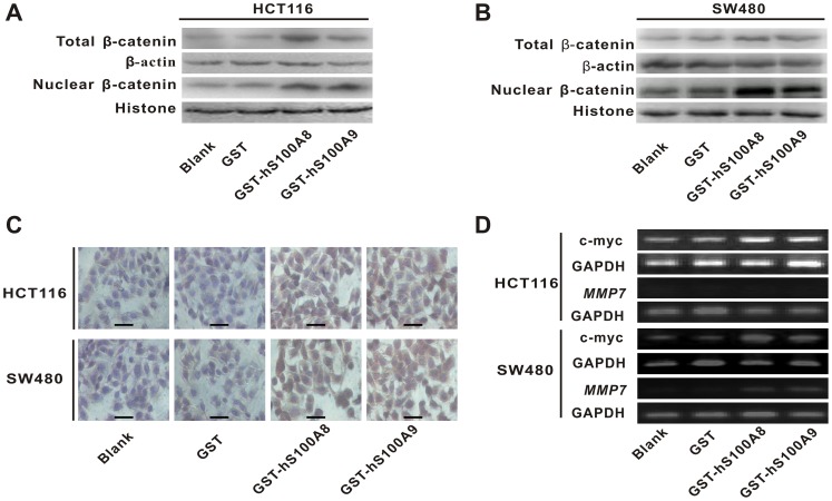
Figure 4. The influence of recombinant S100A8 and S100A9 proteins on the levels of β-catenin and its target gene c-myc and MMP7 in CRC cells.
(A) HCT116 and (B) SW480 cells were treated with and without GST, GST-hS100A8 or GST-hS100A9 at concentration of 10 µg/ml for 36 h, and total and nuclear β-catenin level were measured by Western blot. β-actin and histone were used as internal reference controls. (C) HCT116 and SW480 cells were treated with and without GST, GST-hS100A8 or GST-hS100A9 at concentration of 10 µg/ml for 48 h, and β-catenin level was analyzed by immunocytochemical (ICC) staining. The representative images are shown in the graph. The intense staining for β-catenin level is in nucleus after treatment with GST-hS100A8 and GST-hS100A9. Black scale bars = 100 µm. (D) HCT116 andSW480 cells were treated with and without GST, GST-hS100A8 or GST-hS100A9 at concentration of 10 µg/ml for 48 h, and the expression of c-myc mRNA and MMP7 mRNA were detected using RT-PCR. GAPDH was used as an internal reference control.

