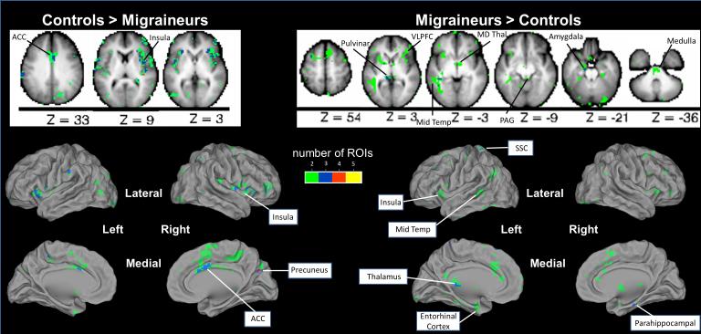
Figure 3. Resting State Functional Connectivity with Pain Regions Differs in Chronic Migraineurs Compared to Controls.
Summary analyses of 2-sample t-tests for each of the 5 pain ROIs identified voxels with rs-fc that differed between chronic migraineurs and controls. Axial slices are shown with the left hemisphere on the left side. Green = the rs-fc of that voxel with 2 of 5 a priori pain ROIs differs between chronic migraineurs and controls; Blue = the rs-fc of that voxel with 3 of 5 a priori pain ROIs differs between groups; Red = the rs-fc of that voxel with 4 of 5 a priori pain ROIs differs between groups; Yellow = the rs-fc of that voxel with 5 of 5 a priori pain ROIs differs between groups. ACC = anterior cingulate cortex; VLPFC = ventrolateral prefrontal cortex; Mid Temp = middle temporal cortex; MD Thal = medial dorsal thalamus; PAG = periaqueductal gray; SSC = somatosensory cortex.

