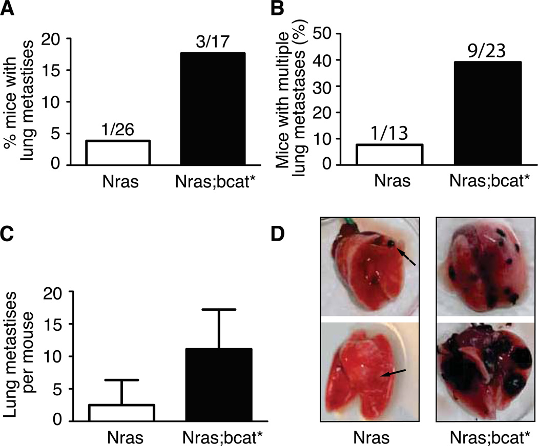
Figure 7. bcat* increases the metastatic potential of murine melanoma.
(A) The lungs of the Nras and Nras;bcat* mice that developed primary melanoma were investigated for distant metastases. (p-value = 0.28, Fisher’s exact test).
(B) The number of nude mice with multiple lung tumours was scored after the injection of 2.5 × 10⁵ cells from Nras or Nras;bcat* tumours into the tail vein. χ2 = 0.043.
(C) Mean number of lung metastases per nude mouse presenting with lung tumours after the injection of Nras or Nras;bcat* melanoma cells into the tail vein (p-value = 0.024).
(D) Lungs from nude mice, following the injection of cells from Nras or Nras;bcat* tumours into the tail vein. Arrows show melanoma tumours. The lungs shown were removed from mice about 100 days after the injection of Nras or Nras;bcat* cells. OCT was injected into the lungs to facilitate the visualisation and preservation of lung tissue.

