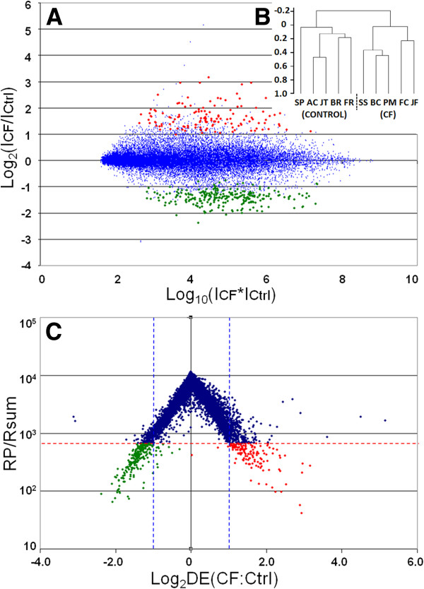
Figure 1.
Visualization of Microarray data. A) R-I plot, in which the log2 of the ratio of mean intensities in CF-vs.-non CF samples is plotted against the log of their product, demonstrating the absence of an intensity dependent fold-change bias. The regulated genes chosen for further analysis using the RP statistic (p < 0.0001 cut-off) are shown in red (up-regulated genes) and green (down-regulated genes). B) Dendrogram of clustered samples, in which CF and non CF samples cluster with respect to their phenotype. C) Volcano plot of microarray data, in which the Log2 of the RP statistic is plotted against Log2 of Fold Change. The p < 0.0001 cut-off (horizontal dotted line) illustrates the gene list chosen for further analysis. Genes outside the vertical dotted lines have more than 2-fold differential expression.

