
Fig. 1.
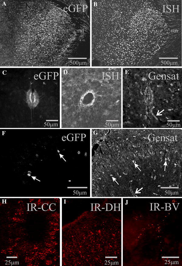
Regional expression of Cx45-eGFP is consistent with in situ hybridisation, reporter expression in another transgenic mouse line and protein localisation. a, c, f eGFP-IR from the Cx45-eGFP mouse. b, d In situ hybridisation images for Cx45, taken from the Allen Brain Atlas. e, g Images of expression in a Cx45 BAC reporter mouse taken from the Gensat project. High levels of expression are evident in neurones in the thalamus (a, b), ependymal cells surrounding the spinal cord central canal (c–e) and in layers I–III in the dorsal horn (f, g). Closed head arrows indicate neurones whilst open head arrows blood vessels apparent due to smooth muscle labelling. Immunoreactivity for Cx45 can also be detected in the ependymal cell layer (h), dorsal horn (i) and in blood vessels (j). b, e, g Inverted from original images to correspond with those shown here. rtn reticular thalamic nucleus. Colour images are available online