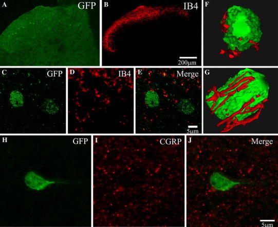
Fig. 4.

a, b IB4 staining is clearly observed within laminae I/II of the DH and makes close appositions to both Cx45-eGFP cells within laminae I/II and those in lamina III. c–e Confocal scan of representative cells (from lamina III in a) demonstrate the density of projections surrounding the Cx45-eGFP cells. f, g A reconstructed model example in two orientations illustrating the density of projections onto the cell soma and putative sites of contact. h–j Staining with CGRP demonstrates that Cx45-eGFP cells do not receive close appositions from CGRP-positive fibres. Colour images are shown online
