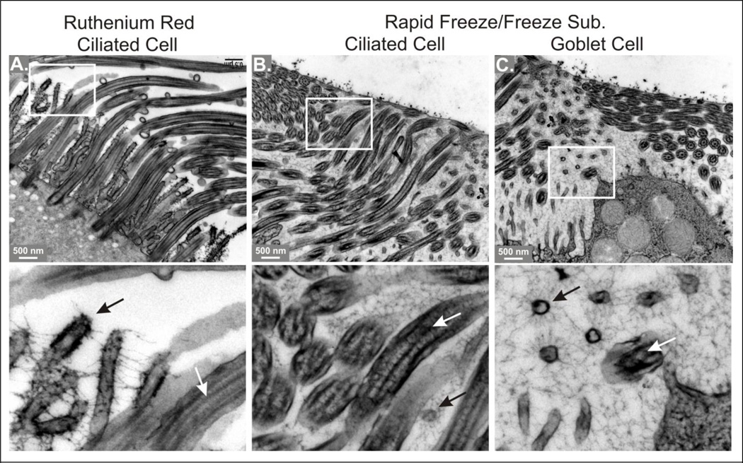
Figure 6. Comparison of fixation methods on mucosal glycocalyx in human bronchial epithelium.
Bottom panels are magnified portions of the images above, as indicated by the white boxes. A. Tissue stained with ruthenium red and processed for conventional EM. Note how ruthenium red causes the glycocalyx to appear as thick, sparse strands, leaving most of the intercellular space apparently open. B and C. Tissue fixed by rapid freezing and freeze substitution, showing areas of the PCL above ciliated and goblet cells, as indicated. Note the finely textured, space filling matrix revealed by RF/FS. Lower panels, black arrows = microvilli, white arrows = cilia. See text for more details.

