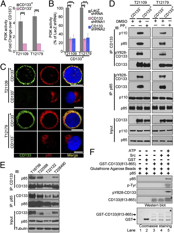
Fig. 2.
CD133 physically interacts with p85. (A) The PI3K activity of CD133+ cells versus CD133− cells derived from glioblastoma specimens were assessed using a PI3 kinase ELISA kit. Values are normalized to that of matched CD133− cells. Results are expressed as mean ± SD from three separate experiments; ***P < 0.001. (B) Targeting CD133 decreases PI3K activity in CD133+ cells. Seventy-two hours after lentivirus infection, the PI3K activity of CD133+ infected with the indicated lentivirus was measured using a PI3 kinase ELISA kit. Values are normalized to that of CD133+ cells treated with LacZ shRNA, which is set as 100%. Results are expressed as mean ± SD from three separate experiments; ***P < 0.001. (C) Colocalization of CD133 and p85 was assessed by immunofluorescence staining of CD133 (green) and p85 (red) in CD133+ and CD133− cells derived from glioblastoma specimens T21109 (Upper) and T12179 (Lower). Nuclei (blue) were counterstained with Hoechst33258. Colocalization of CD133 and p85 is demonstrated by yellow fluorescence. (Scale bars, 10 µm.) (D) Co-IP analysis to determine the interaction between CD133 and p85 in vivo. Lysates of CD133− or CD133+ cells pretreated with DMSO or PP2 (5 µM) for 1 h were subjected to IP using anti-CD133 or anti-p85 antibody, followed by immunoblotting (IB) with anti-CD133, anti-p110, anti-p85, or anti-pY828-CD133 antibodies. Whole-cell lysates were analyzed by IB with anti-CD133, anti-p110, or anti-p85 antibodies as input. (E) Co-IP analysis to determine the interaction between CD133 and p85 in glioblastoma tissues. The lysates of glioblastoma tissues were subjected to immunoprecipitation (IP) using anti-CD133 or anti-p85 antibodies, followed by IB with anti-CD133 or anti-p85 antibodies. (F) In vitro interaction between CD133 and p85. GST or GST-CD133(813-865) (CD133 C-terminal cytoplasmic domain; amino acids 813–865) protein preincubated with or without active Src and ATP in a phosphorylation buffer was incubated with purified p85 protein. The GST pull-down products were blotted with anti-CD133 C-terminal, anti-p85, anti-tyrosine phosphorylation, and anti-pY828-CD133 antibodies (Upper). GST and GST-CD133(813-865) were shown by Coomassie Blue staining (Lower). *Tyr-phosphorylated form of CD133 C-terminal cytoplasmic domain.

