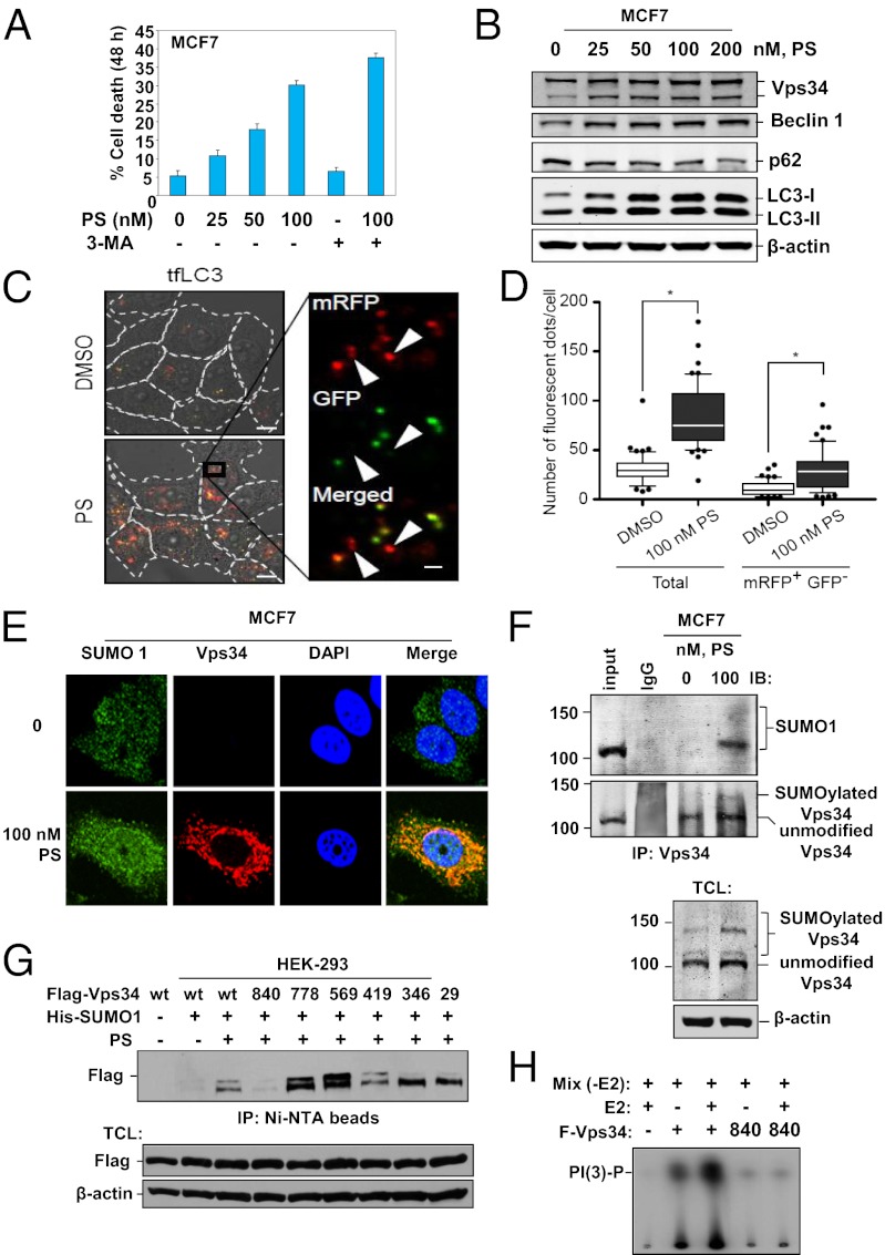
Fig. 1.
HDAC inhibitor panobinostat induces autophagy in MCF7 cells. (A) Cell death of MCF7 cells induced by treatment with the indicated concentrations of PS and/or 3-MA for 48 h. For 3-MA treatment, cells were pretreated with 20 mM 3-MA for 4 h. (B) Depletion of p62 and induction of Vps34, Beclin 1, and LC3 II in MCF7 cells following 48 h of treatment with PS, as indicated. (C) MCF-7 cells were transduced with lentivirus encoding mRFP-GFP-LC3 (tfLC3) for 48 h followed by treatment with 100 nM PS or control DMSO for 24 h. Representative fluorescence images merged with DIC images are shown. Arrowheads indicate mRFP+ GFP− degradive autophagosomes. (Scale bars: 10 µm, main image; 1 µm, magnified image.) (D) The number of mRFP+ GFP+ and mRFP+ GFP− fluorescent dots per cell in C were counted using SlideBook 5.0 software (n = 50) and shown as a box plot. The lines, boxes, and error bars represent median values, 25th–75th percentiles, and 10th–90th percentiles, respectively. Statistical significance was determined using a one-way ANOVA followed by Bonferroni's multiple comparison test (GraphPad PRISM ver. 5.01). The asterisk indicates P < 0.001. (E) Immunostaining of Vps34 (red) colocalized with SUMO1 (green) in PS-treated MCF7 cells. (F) Immunoblot analysis of SUMOylation in Vps34 immunoprecipitates and total cell lysates from PS-treated MCF7 cells. (G) Lysine 840 is a major site of PS-induced SUMOylation of Vps34. Immunoblot analyses of SUMOylated Vps34 in HEK-293 cells cotransfected with wild type or K-to-R mutant Flag–Vps34, His-SUMO1 and treated with 50 nM PS for 24 h. (H) Effect of K840R mutation on in vitro lipid kinase activity of Vps34.

