
Figure 4.
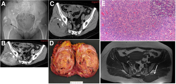
(A) Radiograph demonstrates an ill defined and slightly radiodense area with multiple scattered calcifications of the right buttock (black arrow). (B) Unenhanced CT scan displays a deep soft tissue mass with internal calcifications. (C) The mass is nearly isodense relative to adjacent muscle and contains (mainly in its upper portion (black arrow)) an ill-defined hypodense area that remained nearly unenhanced on post-contrast CT images. Gross specimen (D): The lesion is roundish with polycyclic margins, greyish to yellow in colour, and firm in consistency. Histology (E): Proliferation of bland-appearing spindle cells in a patternless-pattern either in a storiform or haphazard pattern set in a collagen stroma (Hematoxiline & Eosine, 10x magnification). In some areas the cells are arranged around stag-horn haemangiopericytoma-like vessels (Inset, Hematoxiline & Eosine, 20x magnification).