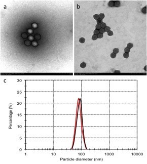
Figure 1.

Particle diameter and TEM photo of DOTAP and cholesterol liposome-encapsulated Ad. The complex of Ad and liposome was visualized by a transmission electron microscope. The naked adenovirus (a) and adenoviral particles encapsulated by DOTAP and cholesterol liposome (b). Both the Ad/Liposome complex and Ad alone shows average particle diameter of the particles, red line represent for Ad alone; black line represent for Ad/Liposome complex (c).
