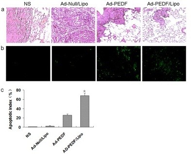
Figure 7.

Histological analysis of tumors. (a) The Hematoxylin and eosin (H&E) staining of lung tissues. After mice were sacrificed histological sections were taken from lungs of mice, and analyzed at × 100 magnification. The mice treated with Ad-PEDF/Liposome showed a significant inhibition of the size of to the metastasis of B16-F10. (b) Detection of apoptotic cancer cells in B16-F10 metastasis in C57 BL/6 mice using TUNEL analysis. The percentage of apoptosis was determined by counting the number of apoptotic cells and dividing by the total number of cancer cells in the field (five high power fields/slide). (c) Percent apoptosis in each group. The group treated with Ad-PEDF/Liposome resulted in significantly increased apoptosis compared to that of other groups (*P < 0.01). Bars, SD; columns, mean.
