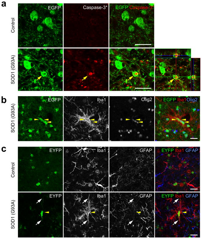
Figure 4.
Apoptosis of oligodendrocytes in the spinal cord of ALS mice. (a) Confocal images from the spinal cord ventral gray matter of a MOBP-EGFP;SOD1 (G93A) mouse at end stage showing an EGFP+ oligodendrocyte (yellow arrow) that was immunopositive for activated caspase-3. Lower right panel is an orthogonal view showing co-localization of activated caspase-3 and EGFP. (b,c) Confocal images of the spinal cord ventral gray matter showing Iba1+ activated microglia surrounding oligodendrocytes labeled with EGFP (b) or EYFP (c) in SOD1 (G93A) expressing MOBP-EGFP (P90) or PLP-CreER;ROSA26-EYFP (P30+60) mice, respectively. Yellow arrowheads highlight several labeled oligodendrocytes, and white arrows in (c) highlight the processes of one EYFP+ oligodendrocyte. Scale bars: 20 μm.

