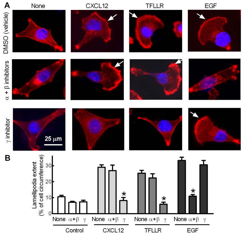
Fig. 4.
Blocking PI3Kγ activity attenuates GRCR-dependent lamellipodia formation of breast cancer cells. MDA-MB-231 cells cultured in serum-free medium were treated with inhibitors of PI3Kα (30 nM) and β (100 nM) or γ (3 μM) 1 h prior to 30 min incubation with CXCL12 (50 ng/ml), TFLLR (25 μM) or EGF (5 ng/ml). Cells were then fixed and subjected to F-actin staining using rhodamine-labeled phalloidin. (A) Images shown are representative of at least 50 cells. Arrows indicates lamellipodial regions. (B) Lamellipodial extent at cell leading edges was quantified as a fraction of cell circumference on 50 randomly selected cells in each group. Columns, means; bars, S.E., *p<0.001 compared to cells without inhibitor treatment (None).

