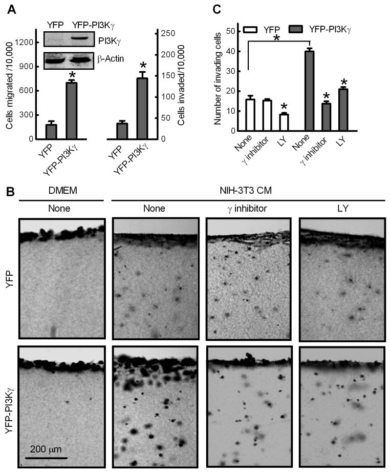
Fig. 5.
Overexpression of PI3Kγ promotes MCF-7 cell migration and invasion. Non-metastatic breast cancer MCF-7 cells were stably transfected with YFP or constitutively membrane-targeted YFP-PI3Kγ. (A) Transwell migration and invasion assays. Data shown are numbers of cells migrated or invaded per 10,000 loaded cells. Bars show the mean ± S.E. (n=5) with *p<0.01 compared to cells transfected with YFP. Inset: Western blot of PI3Kγ and β-actin in MCF-7 cell lines. (B and C) PI3Kγ promotes breast cancer MCF-7 cell invasion of 3D collagen matrices. MCF-7 cells were incubated with type I collagen gels for 48 h in the presence of DMEM or NIH-3T3 CM without (none) or with 10 μM PI3Kγ inhibitor or LY. Gels were fixed, stained and cross-section images were photographed. (B) Images of representative fields, showing the cell monolayer and the underlying collagen matrix containing invading cancer cells. (C) Quantitation of MCF-7 cell invasion. Data are expressed as mean numbers of invading cells per field ± S.E. (n=4) under 10X magnification from ten fields. *p<0.01 compared to cells transfected with YFP or cells in the absence of inhibitors (None).

