
Figure 1.
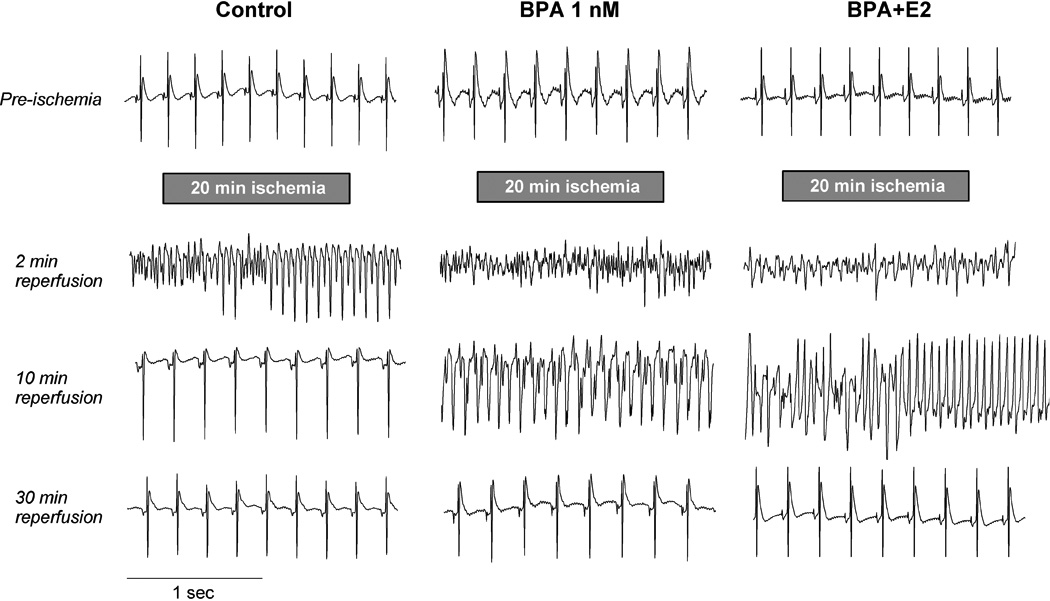
Exposure to low-dose BPA exacerbates ventricular arrhythmia following ischemia/reperfusion in female rat hearts. Shown are typical cardiac electrogram traces recorded from the surface of female rat hearts prior to ischemia, and at various time points (around 2, 10 and 30 minutes) during reperfusion following 20 minutes of global no-flow ischemia. Hearts were exposure to control perfusate, 1 nM BPA or BPA combined with E2 (both at 1 nM) during reperfusion as indicated. All three traces show normal sinus rhythm pre-ischemia. For the control heart, traces show transition from ventricular fibrillation (VF) to ventricular tachycardia (VT) at 2 minutes of reperfusion, and sinus rhythm at 10 and 30 minutes of reperfusion. For the heart exposed to BPA, traces show VF (2 minutes), VT (10 minutes) and sinus rhythm (30 minutes). For the heart exposed to BPA plus E2, traces show VF (2 minutes) followed by transition from VF to VT (10 minutes) and sinus rhythm (30 minutes). It should be noted that a fair amount of variation was observed in terms of the duration of arrhythmias and the nature of arrhythmia at any particular time point; for average data see Figure 2.