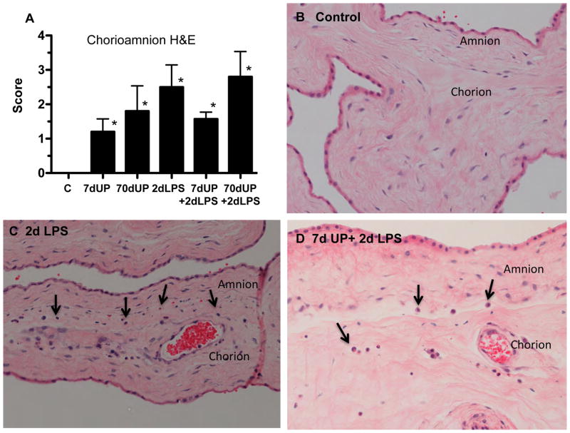
Figure 3. Staging of Chorioamnionitis after intra-amniotic injections of LPS and U. parvum.
(A) Blind scoring for severity of chorioamnionitis was performed using a modified Redline staging method. Representative photomicrographs showing hematoxylin and eosin staining of chorioamnion sections from (B) controls, (C) 2 day LPS, and (D) 7 day U. parvum + 2 day LPS. Arrows point to inflammatory cells at the junction of amnion and chorion (*p< 0.05 versus controls).

