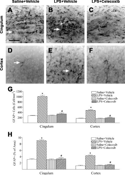
Fig. 6.
Representative photomicrographs of astrocytes (A to C, cingulum area; D to F, cortex) in the rat brain 24 hours (P6) after LPS injection. As shown by GFAP immunostaining in the cingulum white matter (A) and cortex (D), some GFAP positive cells were detected and most of those cells were in resting status with fine processes extending from the main cellular processes (arrows indicated A and D) in the control rat brain. Significantly increased numbers of reactive astrocytes showing hypertrophy of cellular processes of astrocytes (arrows indicated in B and E) were observed in the cingulum white matter (B) and cortex (E) of the rat brain following neonatal LPS exposure. Celecoxib treatment reduced the number of reactive astrocytes stimulated by LPS in the above areas (C, F and G). The scale bar shown in A represents 50 μm for A to F. Quantitation of the number of GFAP positive cells (G) and the percentage area of image that contained GFAP staining (H) in the cingulum white matter and cortex was performed as described in Methods. The results are expressed as the mean ± SEM of six animals in each group, and analyzed by one-way ANOVA. * P < 0.05 represents a significant difference for the LPS + Vehicle group as compared with the Saline + Vehicle group. # P < 0.05 represents a significant difference for the LPS + Celecoxib group as compared with the LPS + Vehicle group.

