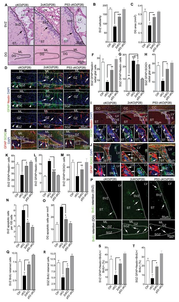
Figure 5. p53 deletion rescues NSCs defects in FIP200hGFAP cKO mice.
(A-C) H&E staining of the SVZ and DG of P28 mice (A). Arrows mark cells in the SVZ. Mean±SE of the SVZ cellularity (B) and DG area (C) per section are shown. (D-M) Immunofluorescence of the DG (D-H) and SVZ (I-M) of P28 mice(D, E, I, and J). The boxed areas are shown in more detail in insets (E) or panels below (I, J). Arrows mark GFAP+/Nestin+ and GFAP+/SOX2+ NSCs with radial glial morphology (D, E), and arrowheads mark GFAP+/Nestin− and GFAP+/SOX2− astrocytes. Mean±SE of the number of GFAP+/Nestin+ and GFAP+/SOX2+ radial glia (F, H), and NSCs (K, M), and GFAP+/Nestin− astrocytes (G, L) per section are shown. (N, O) Mean±SE of the number of TUNEL+ cells per 100 SVZ cells (N) or per 1 mm2 DG area (O) of P28 mice are shown. (P-R) BrdU retention in the SVZ and DG of P31 mice with BrdU+ cells marked by arrows (P). Mean±SE of the number of BrdU+ cells per section are shown in Q and R. (S, T) Mean±SE of percentage of GFAP+/Nestin+/BrdU+ cells of total BrdU retained cells in the SVZ (S) and SGZ (T) of P31 mice are shown. n= 5 mice, ≥4 sections/mouse, >500 cells counted/mouse in N, >20 BrdU+ cells counted/mouse in S and T. For staining panels, lines indicate the boundaries of the SVZ and DG (A) or GZ (D, E, K lower panels). NS: no significance; *: p<0.05; **: p<0.01; ***: p<0.001.

