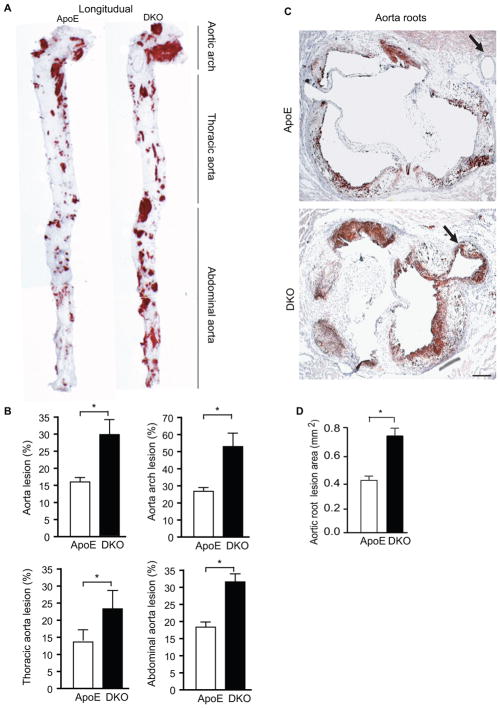
Figure 1. AIP1 deletion facilitates early atherosclerosis progression in ApoE−/− mice.
ApoE−/− and DKO adult mice were fed with Western-type diet for 10 weeks. A–B. Whole aortas were harvested, opened longitudinally followed by en face staining with oil red O. Representative photomicrographs are shown in A with the aortic arch, thoracic aorta and abdominal aorta indicated. Quantifications of atheroma (%) in total and each aortic segment are shown in B. Data are mean ± SEM from 10 mice per group. *, p<0.05, comparing ApoE−/− and DKO groups. C–D. Cross-sections from the aortic roots stained with Oil red-O. Representative photomicrographs are shown in C. Scale bar: 100 μm. Quantification of atheroma area is shown in D. Six sections (150 μm apart for each section) per mouse from 10 mice in each group. Data are presented are mean±SEM, *, p<0.05.

