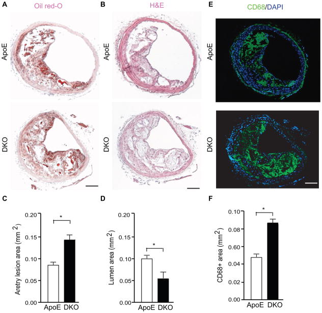
Figure 2. AIP1 deletion increases lesion expansion and macrophage infiltration in the innominate artery in the ApoE−/− mice.
A–C. Representative histological analysis of cross-sections from the brachiocephalic arteries stained with hematoxylin and eosin (H&E), oil red-O, CD68 (a macrophage marker). Scale bar: 50 μm. Quantifications of atheroma area, lumen area and CD68+ area are shown in D–F. Six sections (150 μm apart for each section) per mouse from 5 mice in each group. Data are presented are mean±SEM, *, p<0.05.

