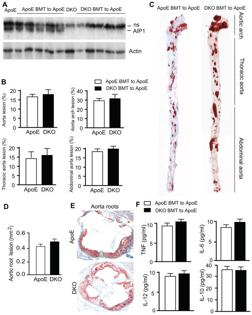
Figure 5. AIP1-KO macrophages do not accelerate atherogenesis and alter inflammatory cytokines in ApoE-KO recipients.
A. Characterization of bone marrow transplantation (BMT). Bone marrow cells from ApoE−/− or DKO mice were transplanted into ApoE−/− recipient mice. Successful BMT was verified in six weeks post-BMT by genotyping the peripheral blood cells for the AIP1 gene (not shown). Peripheral blood cell lysates were also determined for AIP1 protein expression by Western blot with anti-AIP1. Peripheral blood cells from ApoE and DKO mice without BMT were used as controls. AIP1 band is indicated. ns: non-specific bands. B–C. The BMT mice were fed with Western-type diet for 10 weeks, whole aortas were harvested, opened longitudinally followed by en face staining with oil red O. Quantifications of atheroma (%) in total and each aortic segment are shown in B, and representative photomicrographs are shown in C with the aortic arch, thoracic aorta and abdominal aorta indicated. Data are mean±SEM from 8 mice per group. D–E. Cross-sections from the aortic roots stained with Oil red-O. Quantification of atheroma area is shown in D, and representative photomicrographs are shown in E. Six sections (150 μm apart for each section) per mouse from 8 mice in each group. Data are presented are mean±SEM. F. Concentration of cytokines: TNF, IL-6, IL-10, IL-12 in plasma of BMT mice with 10 weeks Western-type diet. n = 8 mice in each group. No significant differences between ApoE and DKO BMT groups.

