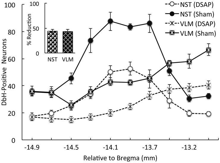
Figure 5.
Anterior vlBST-targeted DSAP lesions reduced the number of DbH-positive NA neurons remaining within the NST-A2/C2 and VLM-A1/C1 cell groups compared to counts in sham-lesioned controls. Inset: Average percent reduction of DbH-positive neurons in DSAP rats (n=18) across ten rostrocaudal levels, relative to counts in sham controls (n=7). The percent reduction of NA neurons in each DSAP rat was calculated using the average counts in sham rats as 100%. Line graph symbols represent group means. Post hoc analyses confirmed that NA cell count differences between DSAP and sham rats were significant at every rostrocaudal level of the NST and VLM (P < 0.05 at each level).

