
Figure 1.
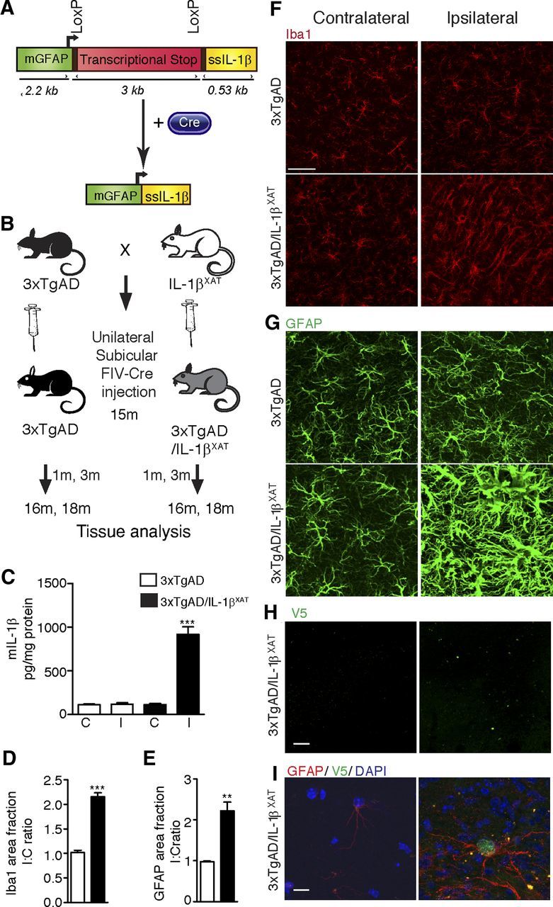
Research design and chronic neuroinflammatory phenotype in 3xTgAD/IL-1βXAT mice. A, The linear construct (∼10 kb) bears the murine GFAP promoter (mGFAP), a transcriptional stop signal flanked by LoxP sites, and the cDNA for hIL-1β gene fused to the signal sequence of mature hIL-1RA (ssIL-1β). After delivery of Cre, the transcriptional stop is excised out and the production of IL-1β is induced locally. B, 3xTgAD mice were bred to the IL-1βXAT mice. Fifteen-month-old 3xTgAD/IL-1βXAT and control littermates of the 3xTgAD genotype from the F1 generation were given a unilateral stereotactic injection of FIV–Cre in the subiculum. Groups of mice were killed 1 and 3 months after stereotactic surgery (at 16 and 18 months of age, respectively), and glial activation as well as amyloid and tau pathology were assessed. C, Murine IL-1β protein ELISA measurements. C, Contralateral; I, ipsilateral. Data were expressed as mean picograms mIL-1β per milligram protein ± SEM per group and analyzed by a one-way ANOVA test, followed by a Tukey's post hoc test. The level of mIL-1β in the ipsilateral hippocampus of 3xTgAD/IL-1βXAT mice is significantly greater than all other groups (***p < 0.0001), but the three other groups are not significantly different from each other (n = 3–9 per group). One month after transgene activation, elevation in markers of glial activation was observed in the ipsilateral subiculum of 3xTgAD/IL-1βXAT mice. D, Area fraction measurements of Iba1 immunohistochemical data. Data expressed as mean I/C ratio ± SEM per group and analyzed by unpaired Student's t test, ***p < 0.0001; n = 5–7 per group. E, Area fraction measurements of GFAP immunohistochemical data. Data expressed as mean I/C ratio ± SEM per group and analyzed by unpaired Student's t test, **p < 0.01; n = 3–4 per group. Representative confocal micrographs of the ipsilateral and contralateral subiculum immunostained with the microglial marker Iba1 (F) and the astrocytic marker GFAP (G) are shown. Scale bars, 30 μm. H, Representative photomicrographs from 16-month-old 3xTgAD/IL-1βXAT mice immunostained with an antibody against V5, a viral epitope tag present in the injected FIV–Cre construct. Scale bars, 200 μm. I, Representative high-power photomicrograph from the same experiment demonstrating the presence of V5 in the nucleus of a GFAP-positive astrocyte in the ipsilateral but not the contralateral subiculum. Scale bars, 10 μm.