
Figure 2.
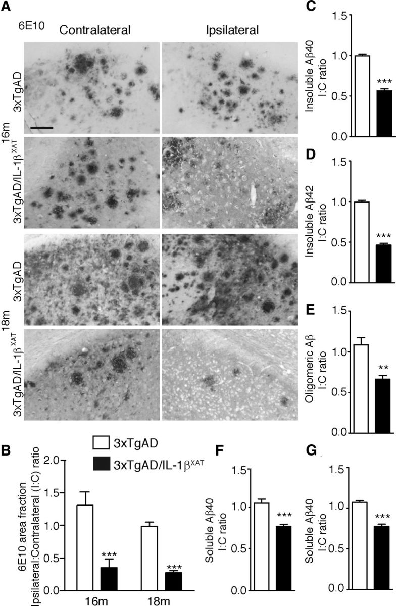
Sustained IL-1β expression ameliorates amyloid load in 3xTgAD/IL-1βXAT mice. A, Representative photomicrographs of contralateral and ipsilateral subicular sections from 3xTgAD and 3xTgAD/IL-1βXAT mice immunostained with the anti-amyloid antibody 6E10 at 16 and 18 months. Scale bars, 100 μm. B, Quantification of 6E10 immunopositive amyloid plaque area fraction in the ipsilateral and contralateral subiculum of 3xTgAD and 3xTgAD/IL-1βXAT mice at 16 and 18 months. Data are represented as the mean I/C ratio ± SEM for each group and was analyzed by a two-way ANOVA with Bonferroni's post hoc test; n = 3–5 mice per group. C–G, ELISA results obtained from whole ipsilateral and/or contralateral hippocampi of 3xTgAD and 3xTgAD/IL-1βXAT mice at 16 months. Mean I/C ratios ± SEM of Aβ40 and Aβ42 in the guanidinium-HCl (Insoluble) fraction (C, D), Tper (Soluble) fraction (F, G), and Oligomeric Aβ in the soluble fraction (E) are shown. In all measures, 3xTgAD/IL-1βXAT mice demonstrated a lower amyloid load in the ipsilateral subiculum/hippocampus at 16 and 18 months compared with 3xTgAD controls; n = 3–9 mice per group. Data were analyzed with unpaired Student's t tests; **p < 0.01, ***p < 0.0001.