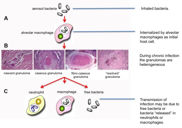
Figure 1. The infection cycle of Mycobacterium tuberculosis.
(A) Infection is initiated by inhalation of infectious bacilli that are likely internalized by alveolar macrophages that patrol the airway surfaces of the lung. Mtb induces a proinflammatory reaction through the activation of TLR and NOD pathways and initiates the formation of a macrophage-centric granuloma. (B) During the chronic or latent phase of infection in humans one can observe a wide variety of granulomas in lung tissue that vary from the productive to the controlled. The phenotype(s) of the productive, infectous granulomas have yet to be formally determined. (C) While transmission is conventionally regarded as cavitation of an infected granuloma(s), there is an increasing body of data arguing that migration of infected neutrophis or macrophages to the airways plays a critical role in the release of bacilli and induction of transmission .

