
Fig. 2.
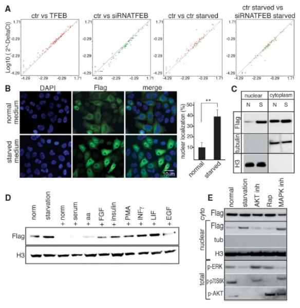
Starvation regulates TFEB nuclear translocation and activity. (A) The expression levels of 51 autophagy-related genes were compared in control and TFEB-overexpressing HeLa cells ultured under different conditions. The results were represented as scatter-plot graphs where circles represent genes with increased (red) or decreased (green) fold change (logarithmic value); x axis, control group; y axis, treated group. (B) Representative images of HeLa cells stably overexpressing TFEB cultured in normal or starved medium for 4 hours. Five fields containing 50 to 100 cells each from five independent experiments were analyzed for TFEB nuclear localization: nucleus, 4′,6′-diamidino-2-phenylindole (DAPI); TFEB, Flag. Values are means ± SEM; Student’s t test (unpaired) **P<0.01. (C) Cells were subjected to nuclear and/or cytosolic fractionation and blotted with antibody against Flag. H3 and tubulin were used as nuclear and cytosolic markers, respectively. (D) Starved cells were treated as indicated (EGF, epidermal growth factor; FGF, fibroblast growth factor; PMA, phorbol 12-myristate 13-acetate). Nuclear fractions were blotted with antibodies against Flag and H3 (loading control). (E) Immunoblot analysis of Flag, tubulin, and H3 in nuclear extracts prepared from normal, starved, and starved then stimulated cells in normal medium for 1 hour (normal) or pretreated with AKT inhibitor, rapamycin mTOR inhibitor, and MAPK inhibitors 1 hour before media stimulation. Total extracts were used to verify the efficiency of the inhibitors (p-ERK, phosphorylated ERK kinase; Rap, rapamycin).