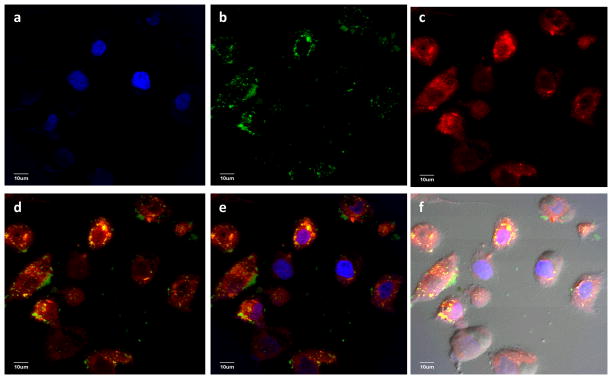
Figure 7.

Laser confocal microscopy images collected for SKOV-3 cell incubated with DOX-HA-SNPs for 24 hours after extensive wash to remove the unbound particles. (a) DAPI channel showing location of nucleus; (b) FITC channel showing location of SNPs; (c) Texas red channel showing location of DOX; (d) an overlay of (b) and (c); (e) an overlay of (a), (b), and (c); (f) An overlay of DIC image and (e). These images clearly demonstrated that DOX-HA-SNPs were internalized inside the cells and not all DOX was released from the NPs as evident from the yellow spots in d–f.
