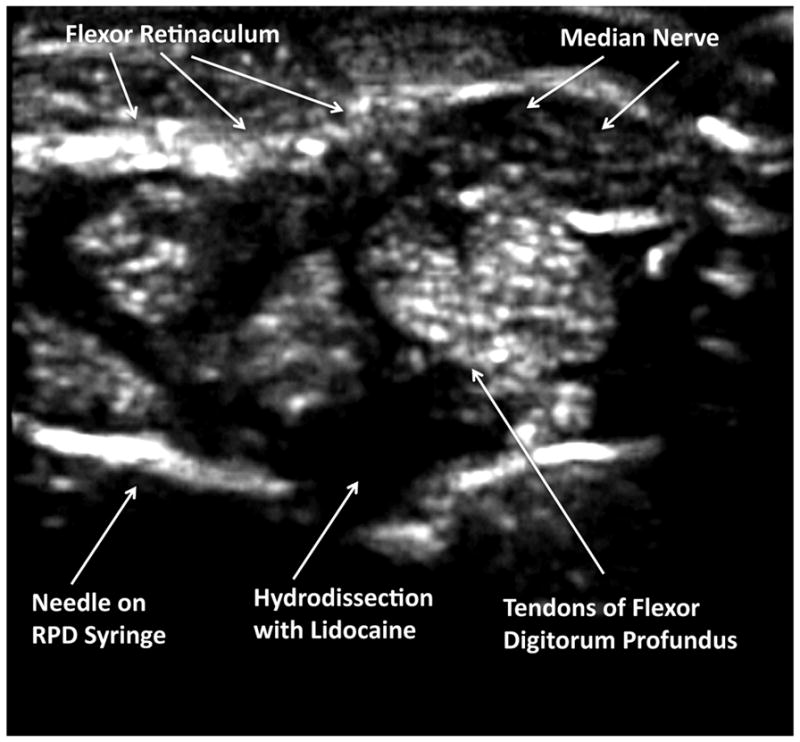
Figure 4. Sonographic Needle Introduction.

In this transverse sonographic image, using the transverse (short-axis) injection approach, the needle is advanced under direct sonographic guidance until the needle tip has pierced the flexor retinaculum, but has not penetrated the fibers of the flexor digitorum profundis. The mechanical syringe is used to determine aspiration of fluid, and then 1% lidocaine is injected to anesthetize the structures and hydrodissect, creating a space for the corticosteroid and lysing adhesions, and pushing away important structures including the median nerve and tendons.
