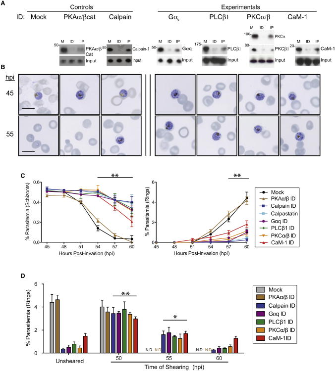
Figure 2. Conserved Function of Host Proteins in P. falciparum-Mediated Cytolysis.
(A) Immunodepletion studies of soluble erythrocyte protein hits confirmed by western blot (M, mock; ID, immunodepletion; IP, elution of target protein immunoprecipitation).
(B) Representative Giemsa images following immunodepletion of host Gαq, PLCβ1, PKCα/PKCβ, and CaM-1 results in persistence of schizont-stage parasites by 60 hpi versus mock or PKAα/PKAβcat depletion, which resulted in exit and reinvasion by 60 hpi.
(C) Flow cytometric quantitation of schizont-stage (left) and ring-stage (right) parasites in depleted erythrocytes. Data shown are means of at least three experiments ± SEM (**p < 0.01).
(D) Ring parasitemia assessed 12 hr following needle shearing of infected and depleted erythrocytes at the times indicated (ND, not determined; **p < 0.01; *p < 0.05). Data shown are means of at least three experiments ± SEM. Also see Figure S2.

