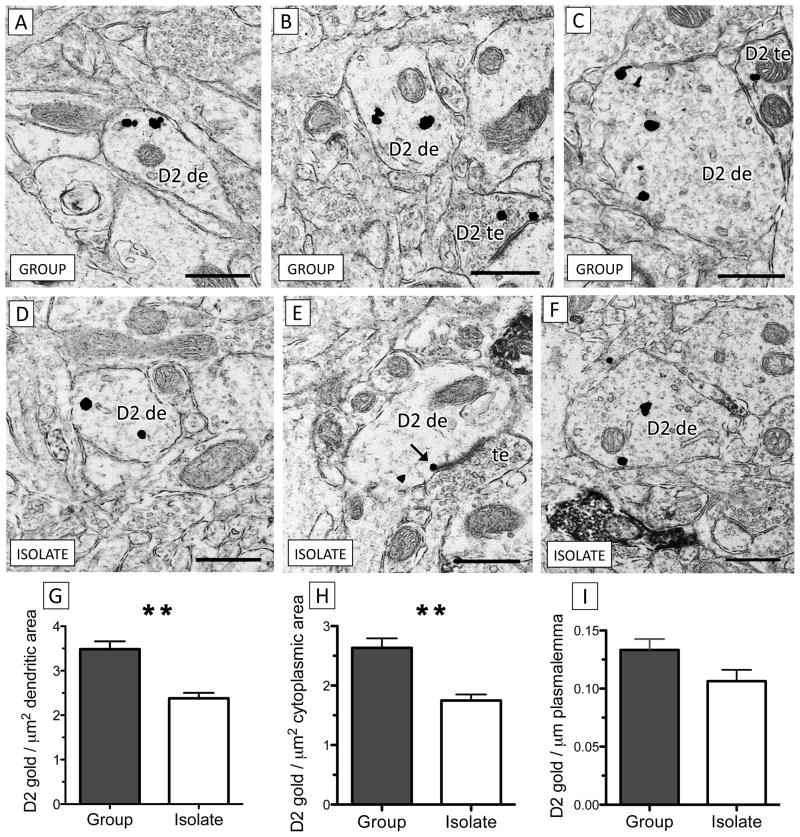
Figure 2.
There is a significant decrease in dendritic D2 receptor expression in the PL of isolated rats relative to group-housed controls. (A–C) Dendrites of various sizes from socially-reared rats contain D2 immunogold (D2 de). In (C), a dendrite containing D2 immunogold is contacted by a D2 terminal (D2 te). (D–) Dendrites from the PL of isolated rats contain qualitatively less D2 immunogold than group-housed controls. In (E), a D2 immunogold particle (black arrow) contacts the outer edge of the postsynaptic density of an asymmetric excitatory-type synapse formed by an unlabeled axon terminal (te). (G–I) Dendrites of isolation-reared rats contain significantly less total D2 (G) and cytoplasmic (H) immunogold per μm2 area relative to group-reared controls but the density of plasmalemmal D2 immunogold particles per μm membrane (I) does not significantly differ between groups (** p < 0.005, n = 9 per group, data is represented as mean ± S.E.M). Scale bars = 500 nm.

