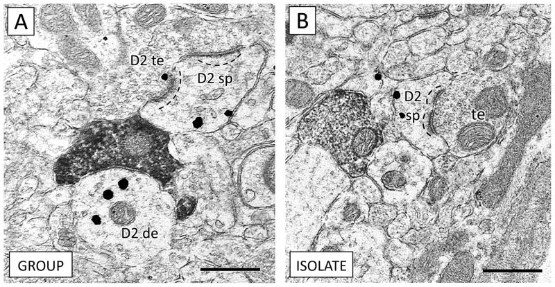
Figure 4.
Expression of dopamine D2 receptors is similar in dendritic spines of group-reared (A) and isolated (B) rats. (A) A D2-containing terminal (D2 te) forms an synapse with a dendritic spine expressing D2 (D2 sp). An apposing CB1 terminal contacts a D2 dendrite (D2 de). (B) An unlabeled terminal (te) forms an asymmetric excitatory-type contact with a spine containing D2 immunogold (D2 sp). In both animals, note the positioning of D2 immunogold opposite the postsynaptic density of the dendritic spines (dashed lines), consistent with dopaminergic input onto spine necks (Freund et al. 1984). Scale bars = 500 nm.

