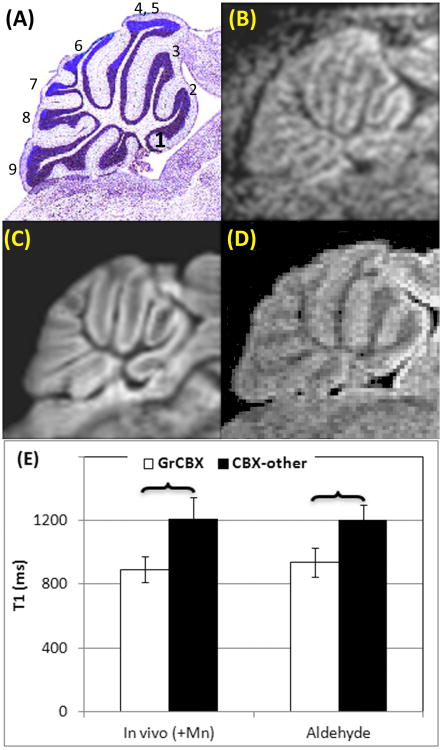
Figure 3.
Sagittal slices of CB. (A): Histology from Paxinos mouse brain atlas. The first to tenth cerebellar lobules were identified and numbered. (B): In vivo MEMRI. (C): Ex vivo MRI of an aldehyde fixed brain. (D): Ex vivo MRI of a brain immersed in MnCl2. (E) T1 values on sub-structures of CBX including GrCBX and other layers measured from in vivo MEMRI (in vivo (+Mn)), ex vivo MRI of brains fixed using aldehyde.
 : significant difference between two structures.
Abbr. in Figure 3: CB – cerebellum, CBX - cerebellar cortex, CBX-other - layers of cerebellar cortex other than granular layer, GrCBX - granular layer of cerebellar cortex

