Figure 5.
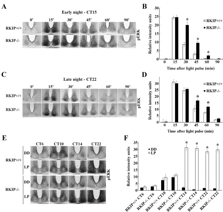
Absence of RKIP prolongs light-induced MAPK/ERK signaling in the SCN but does not alter the gating of photic inputs. A, C, Representative micrographs of p-ERK1/2 expression in the SCN of (top row) RKIP+/+ and (bottom row) RKIP−/− mice at various times (minutes) after a 15 min light pulse is administered at CT 15 (80 lux intensity) (A) or CT 22 (30 lux intensity) (C). Time = 0 represents dark control, whereas samples corresponding to time = 15′ were harvested immediately following termination of the light pulse. B, D, Quantification of light-induced p-ERK1/2 levels in the SCN of RKIP+/+ (white bars) and RKIP−/− (black bars) mice as a function of time (minutes) after the start of a light pulse administered at CT 15 (B) or CT 22 (D). Time = 0 represents dark control. Values are presented as mean ± SEM relative grayscale intensity (in arbitrary units) normalized to background staining. n = 4 mice per group. *p < 0.05 vs RKIP+/+ controls. E, Light-induced ERK1/2 activation in the SCN of RKIP+/+ (top) and RKIP−/− (bottom) mice following a 15 min light pulse (LP: 80 lux) delivered at various circadian times. Single light pulses were given at CT 6, 10, 14 and 22, and mice were killed immediately after cessation of the light pulse. Control mice were killed at the same circadian time without receiving a prior light pulse (DD). F, Quantification of p-ERK1/2 expression in the SCN of light-pulsed (white bars) and dark control (black bars) mice of both genotypes. Values are presented as mean ± SEM relative grayscale intensity (in arbitrary units) normalized to background staining. n = 4 mice per group. *p < 0.05 vs dark control mice of the same genotype.

