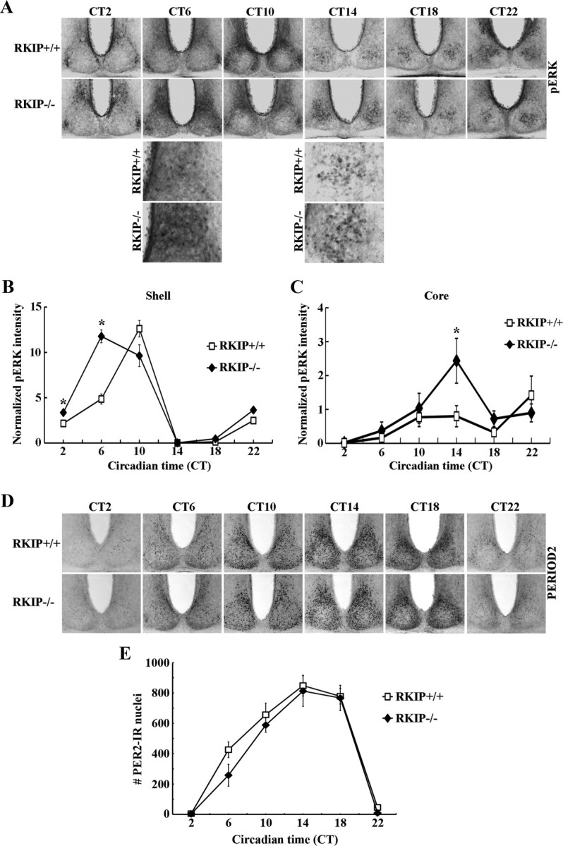
Figure 6.

Absence of RKIP alters the circadian expression profile of p-ERK1/2 in the SCN but does not perturb PER2 rhythms. A, D, Representative micrographs of rhythmic p-ERK1/2 (A) or PER2 (D) expression in the SCN of RKIP+/+ and RKIP−/− mice. Mice were stably entrained to a fixed LD cycle and then released into DD for 2 consecutive days before tissue harvest at the designated circadian times. For (A), higher-magnification images (third and fourth rows) are given to highlight the increase in p-ERK1/2 expression at CT 6 in the shell SCN of RKIP−/− mice, and at CT 14 in the core SCN of RKIP−/− mice, relative to wild-type controls. B, C, Quantification of p-ERK1/2 expression in the shell (B) and core (C) SCN of RKIP+/+ and RKIP−/− mice. Values are presented as mean ± SEM relative grayscale intensity (in arbitrary units) normalized to background staining. n = 4 mice per group. *p < 0.05 vs RKIP+/+ controls. E, Quantification of PER2 expression in the SCN of RKIP+/+ and RKIP−/− mice. Values represent mean ± SEM number of immunoreactive nuclei in the bilateral SCN. n = 4 mice per group.
