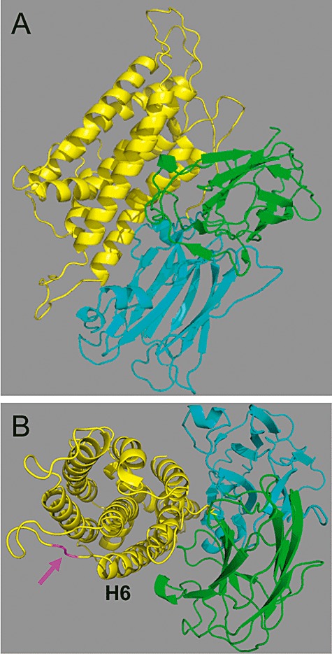
Fig. 1.

Molecular model of Cry48Aa. Model of Cry48Aa showing domain I (yellow), domain II (blue), domain III (green). The cleavage site for Culex gut extracts between the central helices 5 and 6 (H6) is shown in magenta in B. Image created using Pymol software.
